Key factors include:
- The dosage (preferably high-dose region 18-25 Gy single-dose)
- Conformity (maximum dose in the nidus region and minimal dose in the surrounding tissue)
- Selectivity (reduction of the dose in the direct surrounding area of the target volume)
- Accuracy (the radiation exposure and positioning or fixation of the patient)
Effect of the radiation on angiomas / arteriovenous malformations:
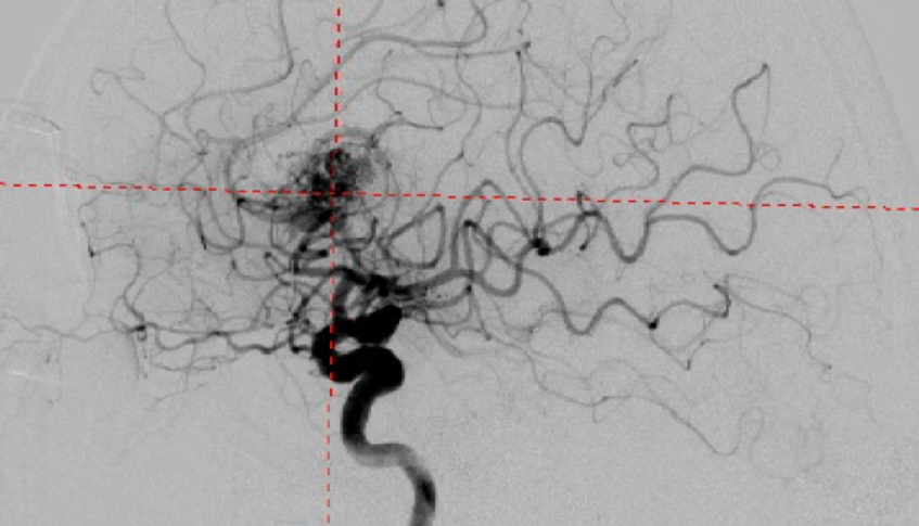
The wall of the blood vessels thickens after irradiation over the course of months to years. This procedure aims to block the changed vessels. This process is called obliteration.
However, the vascular wall changes slowly following radiosurgery and it takes several years (usually 1-3) to completely obliterate the diseased vessel.
When using a combined procedure of radiosurgery and embolization, we recommend performing the radiosurgery first. The embolization can also be additionally performed in the case of incomplete obliteration. If you first perform the embolization however, you have to wait at least 4-6 months due to radiation planning constraints to estimate the recanalization processes.
The probability of obliteration depends on the radiation dose; a higher dose provides better and faster occlusion.
For radiosurgical irradiation, the maximum diameter of the angioma should not be larger than 2.5-3 cm. The radiosurgery should normally target the nidus.
The annual hemorrhaging rate is around 2% after irradiation, the same as the spontaneous hemorrhaging rate.
Effect of radiation on an angioma / arteriovenous malformation:
- High-dose irradiation 20-25 Gy
- Angioma (AVM) should be as small as possible (<2.5 cm)
- Occlusion rate after radiosurgery 70-80%, only apparent after 1-3 years
Aftercare
After radiation therapy we recommend regular MRI checkups. Confirmation of a definite occlusion of the irradiated angioma requires a second angiography after around 1-3 years. If the occlusion was not successful, possible options include surgery, embolization, or repeated radiation therapy.


