How and using what do we treat patients at the Gamma Knife Center?
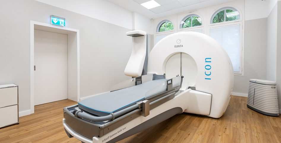
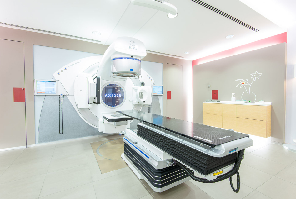
We treat tumors and metastases using radiosurgery with ultra precise devices. We use the Gamma Knife Icon and the stereotactic linear accelerator. Each individual beam from these devices delivers a low dose of radiation. But once these beams converge at a single point the effect is unequaled, similar to how a magnifying glass makes the sun’s rays much stronger. Only at the center of the tumor there is a highly effective beam dose that destroys the source of the disease. This allows us to safeguard surrounding tissue as much as possible with only minimal side effects on the patient. Clinical studies from almost 50 years attest impressively to the treatment’s enormous safety and effectiveness. Pioneered in 1968, this method has been constantly fine-tuned ever since. State-of-the-art computer technology now also makes it even more precise and efficient to use.
Radiosurgery with the Gamma Knife Icon is performed in an outpatient setting or as part of a short hospital stay. Patients do not need to arrive for treatment with an empty stomach. An initial consultation gives us the opportunity to explain the whole process to patients. The efficient, safe procedure allows us to irradiate the source of the disease with an effective dosage. This means a single session is usually sufficient. The treatment itself is completely painless, silent, and lasts between 30 and 120 minutes depending on the application.
Digital imaging devices
The source of the disease is localized just before the treatment. We use the best imaging techniques that allow us to respond quickly to disease changes. We draw on the extensive experience of the neuroradiologists. In addition, we have access to a state-of-the-art imaging infrastructure. For certain diseases, we perform a CT examination in addition to an MRI (magnetic resonance imaging) scan.
Digital subtraction angiography (DSA) in collaboration with the hospital’s neuroradiology department is also used to image blood vessels.
State-of-the-art IT systems (PACS, etc.) transfer all the image data to the Gamma Knife’s planning system.
Treatment planning
We use the acquired image data to demarcate the source of the disease in three dimensions. We overlay all the reference image data acquired with the stereotactic frame to create a precise 3D image. We can also add other image datasets once they have been processed and adapted to the references (e.g. PET examination). These preliminary steps are required to optimally demarcate the source of the disease.
Find out more about the first steps of treatment with the Gamma Knife and cost coverage.
1,213,617 patients were treated with the Gamma Knife worldwide by 2018

Quality management
Since 1998 we have completed over 10,000 successful treatments. Success stories that attest to the outstanding quality and performance of our radiosurgery services.
To constantly meet our own high standards, we introduced an effective quality management system according to ISO 9001 at the outset. The Gamma Knife Center Krefeld and Gamma Knife Center Hanover radiosurgical facilities view quality management as an integral part of service quality and the entire medical treatment process. All the way to outcome monitoring. That is beneficial – for everyone.